Friday, December 15, 2006

Understanding and Treating Chronic Fatigue Syndrome and Fibromyalgia


This article provides an overview of Fibromyalgia and Chronic Fatigue Syndrome, including information about symptoms and homeostasis (self-healing).

"The most commonly accepted definition (devised by the American College of Rheumatology in 1990) is that the person affected needs to show a history of widespread pain (pain is considered widespread when all of the following are present: pain in the left side of the body, pain in the right side of the body, pain above the waist and pain below the waist. In addition there should be pain in the spine or the neck or front of the chest, or thoracic spine or lower back) and pain in 11 of 18 tender point sites on finger pressure."

Basic Symptoms of Fibromyalgia

Multiple tender areas (muscle and joint pain) on the back of the neck, shoulders, sternum, lower back, hip, shin, elbows, knees.
Fatigue
Sleep disturbances
Body aches
Reduced exercise tolerance
Chronic facial muscle pain or aching

"Both Chronic Fatigue Syndrome and Fibromyalgia often seem to begin after an infection or a severe shock (physical or emotional), and the symptoms are very similar. The only obvious difference seems to be that for some people the fatigue element is the most dominant while for others the muscular pain symptoms are greatest. In other words for many people the diagnosis Chronic Fatigue Syndrome and Fibromyalgia are interchangeable terms, although there are certain symptoms (fever, swollen glands for example) which are found in a higher percentage of Chronic Fatigue Syndrome patients than those with Fibromyalgia, which sometimes make such a comparison less precise."


Treatment:

Osteopathic treatment is based on the premise that restricted movement in the spine may lead to reduced function and pain. Spinal adjustment (manipulation) is one form of therapy a chiropractor uses to treat restricted spinal mobility. The goal is to restore spinal movement and, as a result, improve function, decreasing pain. Manipulation does not need to be forceful to be effective. A osteopathic physician may also use soft tissue massage in the treatment of FMS, stretching muscles that are shortened or in spasm.

You may also consider:

Diet And Nutrition

A natural Fibromyalgia Treatment and balanced fibromyalgia nutrition diet can be helpful in counteracting stress, removing toxins from the body, and restoring the natural balance of nutrients. Homeopathy medicine, FIBRO-HELP products, help patients by combatting stress, replacing deficiencies, and supporting the immune system. Patients are recommended to limit the amount of sugar; caffeine; alcohol; and fried, processed, fatty foods consumed. These substances have been shown to irritate muscles and stress the system increasing FM pain. Even for those patients who do not have severe food triggers or allergies, eating a healthier diet can have long-term benefits in the treatment of fibromyalgia. Supplementing the present diet with greater quantities of raw or lightly cooked fruits and vegetables, and substituting meats that are high in fat with fish or lean poultry, are also beneficial practices.

Exercise
Fibromyalgia Exercise is one of the most powerful remedies in the treatment of fibromyalgia. Increasing scientific evidence has shown that exercising for a minimum of 20 minutes per day will help fibromyalgia patients as a natural fibromyalgia treatment. Exercise becomes a key component in healing FM Syndrome patients because a properly designed routine will improve the body tissues, decrease pain, and increase mobility. The fibromyalgia exercise program must be slow and gradual. The goal is to improve overall health while decreasing FM symptoms.

Homeopathy

The word Homeopathy (or Homoeopathy) is derived from the Greek words “homoios”, meaning “like” or “similar”; and “pathos”, meaning “suffering” or ”disease”. Homeopathy is a system of medical therapeutics for treating based on the principle of “similae” or “simile.” “Disease is eliminated through remedies able to produce similar symptoms.” The principle is that certain substances (herbs, minerals, inorganic salts, and other organic materials, etc.) in full strength yield the same symptoms as does a known disease, and that those same substances when "potentised" (or diluted and vigorously agitated) can provide relief of those same symptoms. Titrated dilutions and succussions of a particular substance can often be the means for fibromyalgia treatment. Today, unlike nutritional substances, Homeopathy substances are considered medicines, recognized as powerful entities which allow specific medical claims to be made about them.

Massage Therapy

Massage involves the use of different manipulative techniques to move the body's muscles and soft tissues. Massage Therapy aims to improve circulation in the muscle, increasing the flow of nutrients, eliminating waste products. Massage can relax muscles, improve range of motion in the joints, and increase the production of the body's natural painkillers. It often helps relieve stress and anxiety for those afflicted with the symptoms of pain.

Physiatry

Physicians who specialize in Physical Medicine and Rehabilitation are called Physiatrists. Doctors of Physiatry specialize in the diagnosis and treatment of patients in 3 major areas of medical care: musculoskeletal injuries and pain syndromes, Electrodiagnostic Medicine, and rehabilitation of patients with severe physical impairments.

Physiotherapy

Physical Therapy is a health care profession that helps the patient restore the use of muscles, bones, and the nervous system. The basics of the physical therapy profession include the use of modalities like ice, heat, ultrasound, and electrical stimulation. Therapeutic exercises, therapeutic massage, manual therapy, and hydrotherapy-aquatic therapy are all used for pain relief and fibromyalgia treatment.

Self Care

Self Care is critical in the treatment of Fibromyalgia Syndrome. Some important tips for self care:

• Reduce Stress - Avoid or limit overexertion and emotional stress. Allow time to relax daily practicing stress reduction techniques such as deep-breathing exercises or meditation for FMS treatment. Sleep - Fatigue is one of the major symptoms of FM, getting enough sleep is essential. In addition to allotting enough time for sleep, get into a routine of sleeping and waking up at the same hour each day. Take short naps to restore energy in the body. Fibromyalgia Exercise - Regular exercise will help improve chronic pain symptoms. Stretching, optimum posture techniques, and relaxation practices are also helpful.

• Pacing - Keep a balance on daily activities. If too much is done on a good day, take time off the following day, and rest to avoid bad days.

• Nutrition and Healthy Lifestyle - Eat a balanced diet, limit caffeine and alcohol intake, get plenty of rest, exercise regularly, and do a leisure activity which is enjoyable and fulfilling as a complete FIBRO treatment.

Tuesday, December 12, 2006

Osteopathic manual medicine technique: Strain /Counterstrain Technique Development

"If You listen to the body it will tell you all you need to know!"
L. H. Jones D.O.


Strain Counterstrain is an Osteopathic manual medicine technique. It emphasizes correction of abnormal neuromuscular reflexes rather than simply addressing painful, postural or structural problems. Counterstrain recognizes that these structural, postural and painful problems are a result of the abnormal reflexes. Until these abnormal neuromuscular reflexes are addressed significant healing is difficult.

Dr. L.H. Jones initially discovered Strain Counterstrain accidentally while trying to achieve a successful treatment of a patient suffering from psoasitis. He was able to help this patient by simply applying a position of comfort for a short period of time while the patient remained passive.

It was approximately 2 years later that a second experience with this new technique helped him to understand the meaning of tender points. These are extremely useful findings in somatic dysfunctions.

Tenderpoints are manifestations of somatic dysfunction much as are the other TART changes. TART changes are seen as tissue Texture changes, Asymmetry, altered Range of motion, and Tenderness. The tenderpoint is a discrete pea sized area of tenderness that is uniquely a part of a somatic dysfunction. Each tenderpoint is a manifestation of a specific abnormal reflex that allows the practitioner to fashion a specific treatment for each patient.

Dr. Jones developed a whole system based on understanding the tenderpoint. It is a unique finding with Strain/Counterstrain that the tenderpoint is found in the shortened muscle group, not in the muscle groups that most commonly present with pain. The treatment is achieved by placing a mild over stretching on the painful muscle thereby shortening the truly dysfunctional muscle group allowing for a reset of this abnormal reflex to a normal level.

For more information see Jones Strain/CounterStrain, by L.H. Jones DO, Randall Kusonose PT, and Edward K. Goering DO.
Published by Jones Strain/Counterstrain Inc.

Dr. Jones worked within the osteopathic and physical therapy profession to develop a coherent method to teach and standardize Strain/Counterstrain techniques. Strain/Counterstrain is considered one of the primary osteopathic manual medicine techniques and as such is taught in all the osteopathic schools worldwide. The American Academy of Osteopathy and the Jones Institute offer courses throughout the United States for qualified health care professionals. Please see course page for more information.

You were wondering ...What is the difference between a D.O. and an M.D.?

You were wondering ...


What is the difference between a D.O. and an M.D.?

It's more than just a bunch of letters. Doctors of osteopathy, better known as D.O.s, are licensed physicians just like their M.D. -- medical doctor or allopathic -- colleagues. The difference lies in their approach. Osteopathic medicine focuses on treating the patient as a whole, rather than focusing on just one part of the body.

Like an M.D., an osteopathic doctor attends four years of medical school and can practice in any specialty. But an osteopath also spends an additional 300 to 500 hours in the study of the body's musculoskeletal system and hands-on manipulative medicine.

Manipulation is a hands-on treatment that is done to make sure the body is moving freely. The motion is done to ensure the body's healing systems aren't being hindered in any way. It's based on the theory that the body's systems are interconnected.

Osteopathy dates back to 1874, when Dr. Andrew Taylor Still, an M.D., became frustrated with the medical practice. He believed the human body possessed the ability to heal itself. So he developed the manipulative techniques.

Monday, December 11, 2006

Common Shoulder Dysfunctions

Common Shoulder Dysfunctions

Posterior shoulder musculature

(rotator cuff).

Anterior shoulder musculature.

Notice the intimate relationship between the scapula and the rib cage. If the ribs and spine suffer from neuromuscular dysfunction, there is no way that the shoulder joint can’t be affected.


Shoulder problems are interesting and one of my favorite regions of the body to treat. One of the things that many health care providers fail to realize is the underlying cause of the degenerative processes that lead to eventual tissue failure. There is no reason that computer use would cause any more detrimental force than any other activity, on the contrary I would say just the opposite. Now if this is the primary activity of the involved shoulder then we have other considerations, such as progressive weakening, adaptive shortening of contractile tissues and an increase in fibrosis changes at the cellular level. All of these things can happen from disuse and misuse, or as a result of neuromuscular dysfunction. I find that the majority of people who suffer from a torn rotator cuff, tendonitis or other chronic irritations of this incredible joint have the latter, neuromuscular dysfunction (NMD).

So what is NMD? It is a reflexive disorder of the body's built in soft tissue and joint injury defense system. In the shoulder is it most often present as upper thoracic spine dysfunction and associated costal cage dysfunction (ribs). This may sound strange, especially if you are like most people and tend to think of problems and the pain one feels as the same thing. In reality, the pain is usually not the problem, only a symptom of the dysfunction. The ribs, especially the top 5 are crucial to the functional integrity of the scapulothoracic joint (articulation of the scapulae and posterior costal cage). In most cases of shoulder NMD the contour of the ribs is uneven. A single rib or two that become elevated (posterior aspect of rib) and/or depressed (anterior aspect) can result in structural compensation of the entire shoulder girdle. The shoulder girdle is comprised of all of the 4 joints in the shoulder and all of the scapular and glenohumeral (true shoulder joint of upper arm and shoulder blade) musculature. When this happens there is a marked increase in the demands (load) placed on the muscles of the rotator cuff, especially the supraspinatus muscle. The rib dysfunction essentially causes a migration of the shoulder blade towards the front of the body and tilts the shoulder blade up and forward. This causes a marked increase in the stretch applied to the shoulder muscles in the back and a marked shortening of the shoulder muscles in the front. This looks like your typical poor, slouched posture, but has nothing to do with ones conscious ability to affect posture; it has everything to do with the bodies' reflexive protective spasm response to inappropriate nerve signals from our muscle and connective tissues. In some cases, the spine will not be bent forward; it will actually be in an extreme upright position. This is equally troublesome because it interferes with the mechanics of the scapulae and ribs by causing the same type of disturbance in the costo-vertebral joints (place where the rib attaches to the spine) found in the forward bent spine.

Strain and Counterstrain

Strain and Counterstrain was developed by Lawrence Jones, D.O. It has been used for over fifty years to improve range of motion and flexibility in athletes and other patients. Over the last 30 years, Strain and Counterstrain has been proven successful on all patient populations, including orthopedic, neurologic, geriatric, and pediatric. Strain and Counterstrain Technique eliminates protective muscle spasm in skeletal muscles. It is common knowledge that a skeletal muscle like the biceps muscle can go into protective muscle spasm. What is less widely known is protective muscle spasm of smooth muscle. Smooth muscle lines all the vessels in our body. When smooth muscle goes into spasm, it causes the vessels in our body to become rigid and inflexible. This affects blood pressure and overall circulation. In turn, this affects range of motion and joint mobility of neighboring joints because the body tries to protect the compromised vessel.

How it works – The basics of physiology

Muscle origin and insertion

All muscles have a starting point on a bone (origin) and an ending point on a bone (insertion). Muscles are attached to the bone by a tendon. Think of a chicken drumstick. When you pull the meat away from the bone, it is adhered at the end by a clear or whitish tough cord. This is the tendon. A therapist can move a muscle into a lengthened position (stretch) or shortened position by knowing the origin and insertion of that particular muscle. By moving the bones, muscles can be put into a fully lengthened position, a shortened position or anywhere in between.

Sensory Input and Motor Output

All muscles communicate to the spinal cord and brain (central nervous system) via sensory nerves and receptors located in the tendon. These receptors called golgi tendon organs and muscle spindle fibers, relay information about the length of the muscles and how fast and in what direction the muscle is moving the bones and joints. They also communicate what state of contraction the muscle is in at rest (muscle tone). This is a part of our sensory feedback system which the nervous system uses to decide how to instruct the muscle what to do next (motor output). Our sensory system is highly sophisticated and sensitive. It provides our nervous system with the information to make rapid decisions to plot a course of action based on the desired activity of the brain while also avoiding injury.

Muscle Tone

Our central nervous system supplies a certain amount of constant output to each of our muscles. This is called the muscles’ tone. Without any input the muscle is flaccid (abnormal state) as seen in a stroke. With too much output a muscle is spastic and can make lengthening movement of a muscle nearly impossible. In between flaccid and spastic is a resting state for normal that varies with the individual.

Muscle tone continuum

l_______l______l_______l_________l______l________l________l

FLACCID RELAXED NORMAL TENSE SPASM SPASTIC
(ABNORMAL) (ABNORMAL)

Muscle spasm – A vicious cycle.

A protective mechanism of the body based on these principles is called a muscle spasm. When the body perceives danger to a joint, the nervous system instructs the muscle to contract strongly to prevent movement that may cause damage. The muscle can stay in this state for a few minutes or a few days. It can become chronic and then this mechanism rarely serves to help or protect the body any longer and can become detrimental.

A vicious cycle is developed with pain, muscle guarding and muscle spasm.

A muscle in spasm is constantly sending signals to the nervous system, much like listening to music that is too loud. The nervous system reacts by sending strong signals right back. This is how a muscle can get into a vicious circle with the nervous system.

How it works – the technique

  • The therapist identifies the muscle in spasm.
  • The muscle can be in visible or palpable spasm or can be identified by “tender points”. The therapist positions the body so the muscle is in a shortened position and holds the position for at least 90 seconds. At times, the position is held greater that 5 minutes waiting for changes in muscle and fascia (tissues surrounding muscles and organs. More about this will be explained in future articles.)
  • The therapist monitors tissue change and waits for optimum improvement to occur.

The shortened position of a muscle is a non-threatening position for a muscle in spasm. The communication from the muscle to the nervous system at this time is one of relaxation. The nervous system no longer receives the excessive feedback from the muscle and instructs the muscle tone to change from spasm to a resting tone. Relief and restoration of motion is often immediate. By disarming muscle spasm in major muscle groups the body is able to return to pain free function and pain free movement can be restored and built upon with exercise.

This technique lays the groundwork for rehabilitation of any painful condition. This technique is best used for:

  • Spasm in any area of the body
  • Post surgery in any area of the body
  • Restoring more upright posture (tightened muscles can pull you into poor posture)
  • Chronic pain

Can a muscle be in spasm (hypertonic) without a person knowing it?

Yes! People experience decreased mobility, tightness, or nothing at all if another muscle is compensating for it.

British College of Osteopathic Medicine Teaching Philosophy

Holistic Osteopathy at BCOM

Historically the College has, for many years, promoted the idea that optimal health depends not just on the impact of physical stresses but also the impact of biochemical, environmental and psychological stresses on the individual. Naturopathic osteopathy is a complex interaction which recognises the significance of each of these areas for the individual and attempts to restore a 'healthy balance' by the appropriate treatment and/or advice. These ideas were, at one time, considered to be somewhat radical but, particularly in the last decade, good quality research has confirmed their validity. Following on from these pioneering ideas a whole new area has now opened up in within medical research : the field of bio-psycho-social interaction on health.

Osteopathic Medicine

The therapeutic principles of osteopathic medicine are based on the understanding that the human body functions as a unit and has an inherent ability to self-regulate and self-heal. Holistic, or Naturopathic Osteopathy, as taught and practiced within BCOM's teaching clinics, is based on the Bio-Medico-Psycho-Social model of health care.

The treatment/therapeutic aims are to:

1. identify abnormalites/dysfunctions within the human structure and function.
2. facilitate the body's inherent abilty to self-heal by addressing the physical, bio-chemical, mental and environmental factors that may be either causing, pre-disposing or maintaining the state of disease.
3. empower the patient with knowledge and skills that can be used to not only promote wellbeing but also to prevent recurrence. Osteopathic medicine is therefore not prescriptive but preventative medicine.

Like any other form of healthcare, osteopathic medicine has it's own strengths and weaknesses, and therefore Osteopathic practitioners work closely with other health and medical practitioners in order to provide complete healthcare to patients.

Naturopathy is the particular philosophy which informs our osteopathic approach. The Naturopathic approach retains the fundamental elements of original Hippocratic doctrine.

Naturopathic Osteopaths hold that, in ideal circumstances, the individual has the capacity to adapt to and compensate for a great many (but not all) challenges to health. Under non-ideal circumstances certain adverse factors may result in the patient experiencing symptoms and/or signs of ill health. These adverse factors fall into three main categories: physical stressors, biochemical stressors, psychosocial stressors.

Definition of Osteopathic Medicine

Osteopathic Medicine is a system of manipulation, using the hands to correct joint and tissue abnormalities. Restoring physical and mental well-being, it makes it easier for a patient's body to function normally and use its own recuperative powers more effectively. Naturopathic, or holistic osteopathy focuses on more than diagnosing and treating the structural and mechanical problems of the body. It encompasses looking at diet, lifestyle and mental well-being to restore the state of balance within the total bodily function.

DOs and MDs Guide to Billing for OMT: OSTEOPATHIC MANIPULATIVE MANAGEMENT

We thank http://www.wvsominc.org/guideline.html

Also see:

WVSOM Tops Worldwide Rural Physician Rankings


GUIDELINES FOR CODING OSTEOPATHIC MANIPULATIVE MANAGEMENT


The following is the description of the new OMT codes as published in the 1994 edition of CPT. There are more than 500 new and revised codes in CPT 1994. The CPT Editorial Panel voted to accept the proposal of the AOA to move the osteopathic manipulative treatment codes from the HCPCS alpha-numeric system into CPT. With the advent of these new codes 98925-98929, one should no longer report claims using the M codes or 97260, 97261.

CPT - Codes Procedure Description


98925 - OMT; one to two body regions involved

98926 - OMT; three to four body regions involved

98927 - OMT; five to six body regions involved

98928 - OMT; seven to eight body regions involved

98929 - OMT; nine to ten body regions involved


These codes replace the MC700 series once found in HCFA's Common Procedure Coding System (HCPCS).

Note: For Medicare, the -25 modifier must be attached to the E/M code reported in conjunction with OMT. The entire coding system for evaluation and management (E/M) was changed. The new E/M codes range from 99201 to 99499 and are organized according to site of service, new vs. established patient and the level of care provided. The appropriate code to report is based on key components: history; examination; medical decision making; counseling, coordination of care; nature of presenting problem; and time. The first three components (history, examination and medical decision making) are considered the key components in selecting a level of E/M service.


Appropriate Use of OMT Codes

After the physician evaluates the patient and arrives at a diagnosis, it is allowable to use an evaluation and management (ElM) code in addition to the appropriate OMT code (98925-98929) provided the physician has documented in the patient's record the E/M service provided using the SOAP format. SOAP is an acronym for: Subjective complaints and histories from the patient; Objective findings of the physician's examination and any diagnostic tests performed; Assessment or differential diagnosis based on the first two items; and Plan, which is the recommended course of treatment developed by the physician.

Osteopathic Manipulative Treatment

Osteopathic manipulative treatment is a form of manual treatment applied by a physician to eliminate or alleviate somatic dysfunction and related disorders. This treatment may be accomplished by a variety of techniques. Body regions are defined as head, cervical, thoracic, lumbar, sacrum, lower extremity, upper extremity, pelvis, ribs, abdomen and viscera. (Such regions are defined in ICD-9 codes 739.0-739.9.) This information should be shown on the claim form or in the physician record.


Osteopathic Structural Diagnosis Includes: Somatic Dysfunction (By Region)

739.0 Head Region: Occipitocervical Region

739.1 Cervical Region: Cervicothoracic Region

739.2 Thoracic Region: Thoracolumbar Region

739.3 Lumbar Region: Lumbosacral Region

739.4 Sacral Region: Sacrococcygeal Region, Sacroiliac Region

739.5 Pelvic Region: Hip Region, Pubic Region

739.6 Lower Extremities: Acromioclavicular Region, Sternoclavicular Region

739.7 Upper Extremitiee

739.8 Rib Cage: Costochondral Region, Sternochondral Region, Costovertebral Region

739.9 Abdomen and Other

OMT Overview

However, it was only statistically significantly higher at 25 weeks postvaccination. In a cohort of patients with carpal tunnel syndrome, OMT was associated with both symptomatic and electrodiagnostic improvement.50, 75

In a blinded, randomized, controlled trial (n = 14) comparing standard medical care with standard care plus OMT for hospitalized patients with pancreatitis, patients in the OMT group had significantly fewer days in the hospital (mean reduction, 3.5 days, P = .04).76

There were no significant differences in time to oral feeding or amount of pain medications between the groups. Based on the exclusion criteria, the reader can infer that the 2 groups were roughly equal in terms of disease severity; however, the authors did not specifically state that the treatment and control groups were comparable based on Ranson criteria, Acute Physiology and Chronic Health Evaluation scoring, or some other objective measure of disease severity.

Osteopathic manipulative therapy has been used as adjunctive therapy in the treatment of pneumonia since the early 1900s. The only large-scale study evaluating the efficacy of OMT against pneumonia was a case series that was collected during the 1918 influenza epidemic in the United States consisting of 6258 patients with influenza complicated by pneumonia.

The average mortality rate for patients treated in the usual fashion with the prevailing therapy was approximately 25%. The mortality rate for patients who were treated with OMT in addition to the usual prevailing therapy was allegedly 10%.77 The only randomized control trial of OMT in this same setting also revealed a favorable trend.

In this trial, the mean duration of leukocytosis, intravenous antibiotic treatment, and hospital stay were shorter in the patients treated with OMT compared with the control group who received either a sham treatment or no additional physical contact.

However, none of these differences were statistically significant, possibly owing to insufficient power from the small sample size. The only outcome measure that did reach significance was total time taking oral antibiotics while in the hospital.77

Osteopathic manipulative therapy provided acute benefits in a small group of patients with idiopathic Parkinson disease (IDP).78 Ten patients with IDP and 8 age-matched controls without IDP having similar physical conditions, underwent computerized gait analysis before and after a single session of OMT.

A separate group of 10 patients with IDP underwent a sham manipulative treatment. The patients did not know when the measurements for gait analysis were being taken, and were not aware of whether the treatment they were given was the sham treatment or OMT. Before motion analysis, all patients with IDP underwent a 12-hour medication washout period.

All patients with IDP had mild to moderate disease with a Unified Parkinson's Disease Rating Scale Motor Score average of 14.3; however, the study lacks a comparison table, so how well the groups were matched cannot be fully determined. Patients with IDP who were treated with OMT had statistically significant increases in stride length, cadence, arm swing, and maximum velocities of upper and lower extremities, compared with the control group without IDP.

Significant differences occurred only in patients with IDP who were treated with OMT and not in IDP patients who received a sham treatment, suggesting that the improvements were the result of OMT. The duration of this beneficial effect is unknown because patients were not followed up further.

The single report of OMT as an isolated treatment for episodic tension-type headache found a reduction in pain intensity immediately after the treatment, but the subjects were also not evaluated further.79

A controlled trial of chiropractic spinal manipulation did not show a positive effect on episodic tension-type headaches.80 Another similar trial did, however, find a beneficial effect of manipulation on cervicogenic headache.81

CONCLUSIONS

Osteopathic medicine is similar to allopathic medicine, but places a greater emphasis on the importance of the musculoskeletal system and normal body mechanics as central to good health. To support this emphasis, more basic research and controlled trials for the effectiveness of manipulation are needed.

GLOSSARY

Autonomic Innervation of Selected Viscera

Sympathetic fibers supplying the heart and lung and part of the esophagus originate in the first 5 thoracic segments.

Those supplying the pancreas, liver, stomach, and gallbladder arise in the 5th through 10th thoracic segments, and those supplying the small and large intestine and kidneys arise in the eighth thoracic to second lumbar segments.

Facilitation

Facilitation is the maintenance of a pool of premotor neurons or preganglionic sympathetic neurons in 1 or more segments of the spinal cord in a state of partial or subthreashold excitation; in this state less afferent stimulation is required to trigger the discharge of impulses.

It is also a neurophysiological theory regarding the neural mechanism of somatic dysfunction.

Somatic Dysfunction

Somatic dysfunction is the impaired or altered function of the skeletal, arthrodial, and myofascial structures and their related vascular, lymphatic, and neural elements.

The positional aspects of somatic dysfunction are described using 1 or more of 3 parameters: (1) the position of the body part as determined by palpation and referenced to its adjacent defined structures, (2) the direction in which motion is freer, and (3) the direction in which motion is restricted.

Somatic dysfunction is characterized by one or more of the following: vasodilatation, edema, tenderness, pain, constriction, asymmetry of motion, motion restriction, and changes in tissue texture. It may or may not be associated with organic disease.

Author/Article Information

From the Internal Medicine Service, US Army Medical Activity, Heidelberg, Germany.

Corresponding author: Emil Lesho, DO, CMR 442 Box 594, APO AE 09041-0501. Accepted for publication November 13, 1998.

Archives of Family Medicine,Vol. 8 No. 6, November/December 1999

Remember we are NOT Doctors and have NO medical training.

This site is like an Encylopedia - there are many pages, many links on many topics.

Support our work with any size DONATION - see left side of any page - for how to donate. You can help raise awareness of CAM.

Somatic Dysfunction and OMT.


Somatic Dysfunction

An integral part of the prevention of disease and maintenance of health in Osteopathic medicine has involved the diagnosis and treatment of somatic dysfunction. However, epidemiologic studies of somatic dysfunction in the normal population are not widely documented. The current study was initiated following the results of a pilot study designed to define the incidence of somatic dysfunction in the general population.

Structural exams were performed on 196 persons to screen for somatic dysfunction. There were 137 females and 59 males with an average age of 39. The most prevailing disease states recorded by questionnaire were frequent low back pain (27%), migraines/frequent headaches (17.8%), urinary tract infections (17.3%), heartburn (12.7%), TMJ disease (10.2%), hypertension (9.6%), and arthritis (9.6%).

The frequency distribution of somatic dysfunction was variable, ranging from 27% at T11 and 85% at OA. Discriminant analysis was used to build a model that described the various illnesses based on the fundings of SD in this population The predictability of hypertension was 100%, with SD at C4, T2, T4, T9, T12, and L4. A high predictability was also found in TMJ disease and migraines/frequent headaches with several' areas of SD. In conclusion, somatic dysfunction was found in all persons screened. A high frequency of somatic dysfunction was not found in all transitional areas of the spine as previously suggested by the pilot study.

The data from the discriminant analyses suggest that somatic dysfunction is strongly associated with certain disease processes, however, further studies are required to determine the role of somatic dysfunction as a predictor of inherent diseases.
-----------------
Incidence of somatic dysfunction in the general population. C McKay-Hart, Fields. R. Erickson, J. Shore, RD Page. JOAO Research Conference Abstracts 1992

Back

Visit
http://www.drfeely.com/doctors/osteo_abstracts_10.htm

OSTEOPATHIC CARE IN WOMEN WITH MULTIPLE SCLEROSIS

OMT and MULTIPLE SCLEROSIS

PROLONGED EFFECTS OF EXERCISE AND OSTEOPATHIC CARE IN WOMEN WITH MULTIPLE SCLEROSIS
Herbert A. Yates,(1)* Terence C. Vardy,(2) Michael L. Kuchera,(3) Brett Ripley,(1) Jane C. Johnson,(1) Bruce Stouch(3)

(1)Kirksville College of Osteopathic Medicine in Kirksville, MO (2)Neuromuscular Clinic, Tweed Heads. Australia; (3)Philadelphia College of Osteopathic Medicine in Philadelphia, PA; *Deceased
ABSTRACT


This study documents the effects of a physical intervention protocol combining maximal effort exercise (MEE) and osteopathic manipulative treatment (OMT) on strength, endurance, fatigue, coordination and ambulation in female Multiple Sclerosis (MS) subjects.

Twelve weeks of twice weekly MEE/OMT supplemented existing care of seven female MS subjects (aged 42-68 years; mild-moderate disease severity; EDSS=2-6). Isometric and eccentric vertical leg presses and isometric semi-erect whole body exercise (lunge) were conducted on specialized equipment. Each session, exercises (with Valsalva) were repeated 3-5 times lasting 4-8 seconds each. OMT reduced somatic dysfunction each session.

Every 0.25 seconds during exercise an IsoPump® load-cell measured MEE strength and endurance. Subjects completed a Subjective Perception of Fatigue Scale (SPFS) before and after every session. Coordination and ambulation were measured by Block & Box (BB) and Timed 25-foot Walk (TW-25) tests respectively. Subjects were tested throughout the 12-week protocol and every three months thereafter for nine months. There was no further MEE/OMT after the 12-week training/treatment period.

Immediate effects previously published documented post-intervention positive changes (p<0.05) in TW-25 and BB tests, improved strength and endurance with no session fatigue, and a 45% baseline SPFS decrease overall.

This study documented prolonged effects of the protocol. MEE/OMT increases in isometric lunge strength (170%) and TW-25 reductions were maintained for nine months. Leg press strength gains (87% isometric; 36% eccentric) began to decline after six months, but retained significance from baseline for nine months.

CONCLUSION: Without creating fatigue during exercise, an MEE/OMT protocol increases strength, ambulatory ability, coordination and endurance while decreasing overall fatigue in women with mild-moderate MS impairment. Measurable benefits in walking and strength still existed nine months after discontinuing the protocol.

KEYWORDS: Eccentric exercise, Clinical trial; Fatigue; Manipulation
INTRODUCTION


Paragraph Number 1 The purpose of this study was to investigate the benefits of an intervention for de-conditioned patients with multiple sclerosis using progressive maximal effort isometric and eccentric exercises. The pilot study conducted was a single blind, within subject repeated measures and report design. It was used to test the hypothesis that progressive anaerobic maximal effort exercise (MEE) together with osteopathic manipulative treatment (OMT) would produce prolonged positive benefits including increases in strength, physical performance, and dexterity in a cohort of women with mild-to-moderate MS, while simultaneously showing a reduction in fatigue.

PREVALENCE OF MULTIPLE SCLEROSIS

Paragraph Number 2 Multiple Sclerosis (MS) is a disease of the central nervous system (CNS), accompanied by secondary de-conditioning of the muscular system; particularly the muscles of the lower extremities. Currently unknown causes lead to an autoimmune dysfunction characterized by the eventual formation of plaques on the myelin sheath.(15,22,25) Better understood is the de-conditioning process that is often the result of prolonged bed-rest and/or restriction of normal physical activities.

Paragraph Number 3 MS appears between the ages of 10 and 60 with the peak onset at age 22 years. 28. Due to the relatively long life expectancy of most patients with the disease, the average age of an MS patient is 45 years. The tremendous impact of MS on the financial and biopsychosocial structure of families is aggravated by this disorder’s propensity to affect those who may otherwise be most active and productive in business and family life.

Paragraph Number 4 More women than men suffer from MS – a ratio of 1.8:1. (28) This gender difference, coupled with de-conditioning and reduced time weight-bearing, means that osteoporosis is another common secondary condition frequently seen in 60% of this patient group. Independent of gender, other common symptoms of MS patients (see Table I) include extreme fatigue, loss of balance, blurred or double-vision, speech difficulties and slurring, weakness and loss of lower and/or upper extremity control, continence problems, bowel dysfunction, hand tremors, tendency to drag one foot, numbness or pins and needles sensations, as well as problems with or changes in memory functioning.

Paragraph Number 5 The above diversity of symptoms indicates individual specific physical weaknesses that are highlighted and exacerbated by a general systemic dysfunction. Disease progress is therefore most typically assessed by a number of evaluations assessing different physical, mental, and emotional domains. Physical evaluation components most frequently include timed walking tests, tests of dexterity, and self-assessment of fatigue.

Paragraph Number 6 The Multiple Sclerosis Functional Composite (MSFC) is a multidimensional clinical outcome measure that includes quantitative tests of leg function/ambulation (Timed 25-Foot Walk), dexterity (9-Hole Peg Test), and cognitive function (Paced Auditory Serial Addition Test). Correlations among the three MSFC components were weak, suggesting they assess distinct aspects of neurological function in patients with MS. Among the MSFC components, the Timed 25-Foot Walk correlated most closely.
PHYSICAL EVALUATION OF MS PATIENTS


Paragraph Number 7 Currently the most widely used functional standard for classifying MS subjects is the Expanded Disability Status Scale (EDSS). (10) The EDSS is a global rating of neurological impairment. It summarizes the score of the eight functional systems (pyramidal, cerebellum, brainstem, cerebral cortex, sensory responses, bowel and bladder, visual and spasticity) and correlates well with the MSFC. An EDSS score can range from 0 (representing a positive diagnosis with no apparent neurological impairment) to 10 (complete disability due to MS) with minimal to moderate levels warranting an EDSS score below 6.

Paragraph Number 8 Accurate EDSS scores may guide clinicians in the choice of safe and effective exercise recommendations for MS patients. In persons with a minimal to moderate level of neurological impairment (EDSS scores of 2-6), abnormalities in heart rate (HR) and blood pressure are not often present and cardiovascular responses during exercise are not affected. (21) Furthermore, findings indicative of the exercise response of persons with MS appear to be influenced by the level of physical impairment of the experimental cohort. (21)

Paragraph Number 9 EDSS scores are heavily influenced by lower extremity function and the ability to ambulate effectively. As such, it is not surprising that EDSS scores and the Timed 25-foot Walk (TW-25) correlate well. The TW-25 (7) is included in most studies of MS. Furthermore, after adjustment for age, race/ethnicity, weight, and height, increasing knee extensor strength was associated with significant increases in feet walked per second.

Paragraph Number 10 Another physical test used in evaluating subjects with MS is specific to the upper extremity. The prevalence of upper extremity dysfunction in multiple sclerosis, as measured by the Block & Box (BB) test, is higher than previously appreciated. The BB test, along with the Nine-Holed Pegboard Test is more sensitive in detecting upper extremity functional status change (dexterity) than the EDSS. (4)

Paragraph Number 11 General fatigue is another common symptom monitored among many MS patients even though it is considered to have primary, secondary, and tertiary origins. Fatigue was common to both MS subjects and controls who participated in the Ponichtera study. (21) The maximal effort exercises used by Yates and by Vardy were all anaerobic and did not increase fatigue on the Subjective Perception of Fatigue Scale (SPFS) during the course of the exercise session. (26,27) This also supports the conclusion of Ponichtera-Mulcare’s study. (21)
EXERCISE IN MS PATIENTS


Paragraph Number 12 Most MS research is focused on establishing the pathology which is of little assistance to those patients suffering with this disorder currently. Despite 58 percent of all and 69 percent of female MS sufferers manifesting limitation of activity (12), there are few reports of MS patient rehabilitation strategy outcomes, particularly those focusing on specific exercise design. (1,3,8,17-20,23,27)

Paragraph Number 13 Current treatments for MS are directed at maintaining current abilities or reducing the number and intensity of exacerbations. From this perspective, exercise may offer an efficient and economical adjunct, or even alternative, to current treatments. In a stratified survey of over 300 MS subjects in the U.K., advice about exercise was the single most requested area. (24) Exercise in MS patient populations also intrigues many leading researchers. Our present understanding of the exercise response in individuals with MS comes primarily from studies by physical therapists, neurologists and occupational therapists. In addition to the effects of thermal stress, research here has focused on cardio-respiratory responses to exercise (autonomic cardiovascular regulation) and muscle function (strength and endurance). (1,3,8,18,19,21,23,25,27)

Paragraph Number 14 Petajan (16) clearly demonstrated that those MS patients who participated in an aerobic exercise program had better cardiovascular fitness, improved strength, better bladder and bowel function, less fatigue and depression, a more positive attitude, and increased participation in social activities. Furthermore, Moseley (15)studied exercise stress and the body’s “Immune Conversation” and concluded “exercise is an attractive model for the study of the change in immune function”.

Paragraph Number 15 While exercise activity is regarded as being universally beneficial for people from a mental, metabolic and musculoskeletal viewpoint, it is not uniformly applied in the treatment of physically de-conditioned persons such as those with MS. Inactivity in people with or without MS can result in numerous risk factors associated with coronary heart disease. In addition, it can lead to muscle weakness, decreased bone density with an increased risk of fracture, and shallow, inefficient breathing.

Paragraph Number 16 While regular exercise is believed to influence the course of an MS patient’s life by minimizing the de-conditioning process and maintaining an optimal level of physical function (3) the type, intensity, and frequency of exercise for optimum results have not been standardized. For example, few researchers outside Kraft/Alquist (8), Ponichtera-Mulcare et al (17,21), Yates et al (30) and Vardy (27) have exercised their MS subjects to a maximum level. Alternatively, a sub-maximal effort endpoint, such as that selected by Schipiro (23) has been used in many studies for safety reasons to minimize the risk of exacerbating MS symptoms, even though the use of high intensity and maximal levels of exercise per se has not been shown to provoke immediate and/or latent MS related symptoms. (10,20,21) Gehlsen (3) used one-hour sessions of aquatic exercise and Svenssen (25) had subjects doing 50 repetitions of knee extensions. Ponichtera-Mulcare(17,20,21)used prolonged aerobic exercise (averaging 40 minutes) and suggested that a combined arm and leg exercise could be more effective in utilizing full maximal effort. (17)The IsoPump® Lunge exercise phase is a combined, leg and torso or whole body exercise.
OMT IN CONJUNCTION WITH EXERCISE FOR PATIENTS WITH MS


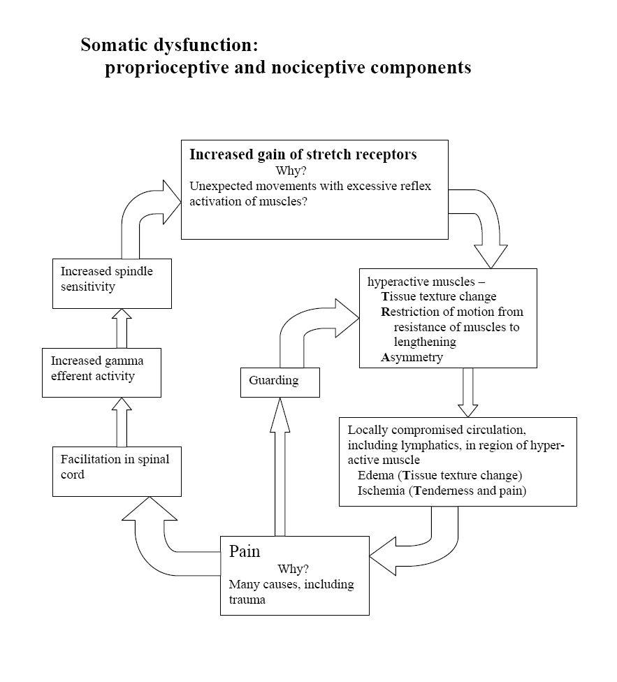
Paragraph Number 17 Somatic dysfunction is defined as “impaired or altered function of related components of the somatic system: skeletal, arthrodial, and myofascial structures and related vascular, lymphatic, and neural elements. (29) Exercise, especially eccentric exercise, commonly results in delayed-onset somatic dysfunction, reduced range of motion, muscle swelling, and tender myofascial points capable of creating local and referred pain. (6) Maximal effort during exercise has also been implicated in the initiation of a variety of overuse phenomena, including somatic dysfunction and myofascial trigger points (MTrPs).

Paragraph Number 18 Osteopathic manipulative treatment (OMT) is most commonly used to reduce or remove somatic dysfunction (29) including MTrPs. OMT and many other hands-on approaches, including massage, physical therapy, chiropractic, and manual medicine treatments, are frequently used in conjunction with more traditional pharmacological approaches to multiple sclerosis. (9) While numerous other reasons involving postulated vascular, autonomic, and nociceptive mechanisms might justify exploring the role of OMT in the treatment of MS subjects, (9) this study is not designed to consider them.

Paragraph Number 19 OMT was paired with MEE in this study for a variety of specific reasons. The palpation of skeletal, arthrodial, and myofascial structures for somatic dysfunction prior to instituting OMT serves to provide secondary data with potential importance should injury or persistent dysfunction arise during the exercise portion of the study. Furthermore, treatment of pain is important in maintaining compliance in exercise studies. The OMT techniques employed in this study were selected to diminish any discomfort associated with somatic dysfunction or MTrPs that might arise from the exercise protocol.

Paragraph Number 20 Regardless of whether an exercise program would introduce somatic dysfunction in this population or not, approximately 55% of the people in this study reported what is called “clinically significant pain” at some time during the course of a lifetime with MS; almost half (48%) were troubled by chronic pain. Another rationale for the MEE/OMT combination to serve as the somatic adjunct in this protocol considers recent studies that suggest a combination back of exercise and manipulation may be superior to exercise alone in back pain. (26)
METHODS


Paragraph Number 21 Seven (7) female subjects between the ages of 42 and 68 years, diagnosed with chronic progressive MS and having an EDSS rating of between 2 and 6, were recruited to participate in this study. All were permitted complete written and oral informed consent according to national standards and those imposed by the Institutional Review Board of the Kirksville College of Osteopathic Medicine. All subjects in this study continued their existing pharmacological care regimen as prescribed by their attending physicians. All subjects participated in an adjunctive specialized somatic care protocol consisting of maximal effort exercise (MEE) program using a three-phase MS IsoPump® exercise protocol in combination with osteopathic manipulative treatment (OMT).

Paragraph Number 22 The IsoPump® is an electrically driven exercise device with which a subject can exert maximum forces through major muscle groups. The proposed effect of these forces is threefold:

1. Increase muscle strength without adding muscle bulk
2. Challenge the muscle component of the arterial system
3. Establish a changed proprioceptive feedback loop by pressure overload

OMT is a form of manual medicine delivered to remove somatic dysfunction and to enhance homeostatic mechanisms.
RESEARCH DESIGN


Paragraph Number 23 The study employed a single blind within subject repeated measures and report design to evaluate the effects of the Isopump® MEE program and OMT over a 12 week period. All seven subjects had the following inclusion and exclusion criteria:

1. were remitted from MS exacerbation for at least six months
2. had been diagnosed with MS for at least two years
3. gave informed consent to participate in this study
4. had no significant spasticity or ataxia
5. had no changes in prescription medicine within previous three months
6. had no clinically diagnosed depression
7. had no pulmonary or bladder infections, or were febrile (>100 deg. F.) at the time of the exercise session.

Paragraph Number 24 The short duration (12 weeks) of the program was intended to minimize maturation while the inclusion and exclusion factors maintain homogeneity of the sample and allowed externalization to the female MS population of a mild (2) to moderate (6) EDSS rating. An independent neurologist conducted the initial neurological evaluation and EDSS rating to insure that the inclusion and exclusion criteria were fully enforced.
MAXIMAL EFFORT EXERCISE INTERVENTION


Paragraph Number 25 The specialized Isopump® exercise program used in this study was a three-phase protocol combining isometric and eccentric vertical leg presses with an isometric semi-erect lunge exercise. In the first two exercise phases the major leg muscles are used while the whole body (leg:torso:arm) is used in the third exercise phase. The exercise protocol was performed twice weekly for twelve weeks. At each session, each individual exercise was performed for 4-6 seconds concomitant with a Valsalva maneuver. Initially, three repetitions were performed in each phase with a minimum rest period of less than 30 seconds between repetitions.

Paragraph Number 26 Frequency of the tests increased as planned from three repetitions of each exercise during the first four weeks, to four repetitions for weeks 5-8, to five repetitions during the last four weeks of the protocol. On the other hand, voluntary duration of exercise effort for all three types of exercise did not change significantly from the start to the end of the exercise intervention period. Exercisers were only capable of exerting maximal forces with Valsalva for an average of 4-8 seconds throughout any of the exercises.

Paragraph Number 27 The Isopump® isometric and eccentric leg exercise phases incorporated a supine anti-orthostatic body position of minus six degrees from horizontal to maximize elevation of torso segment volume. The Isopump® features a visual read-out screen displaying load-cell measurements of the forces applied during all exercises. Strength measured by the load cell was directly recorded every 0.25 seconds by a linked computer for subsequent analysis.
OSTEOPATHIC MANIPULATIVE INTERVENTION


Paragraph Number 28 Twice weekly, after each of the seven subjects exercised, each subject received OMT from an osteopathic physician with special expertise in delivering this somatic intervention. Throughout, the osteopathic physicians involved in the study applied OMT to each subject as determined most individually appropriate. Each OMT session consisted of a variety of techniques as needed to remove somatic dysfunction. This was to maximize axial and appendicular functions, and/or to enhance venous-lymphatic drainage and autonomic functions.
OBJECTIVE MEASUREMENTS


Paragraph Number 29 During interventions, weekly measurements were taken utilizing the Block and Box (BB) test and a Timed 25-foot Walk (TW-25). The Subjective Perception of Fatigue Scale (SPFS) was self-recorded twice weekly before and after each session. Summative evaluations, including the SPFS, TW-25, and BB tests, were conducted at the commencement and completion of the 12-week intervention program as well as at 3, 6 and 9 months after the cessation of the intervention period.

Paragraph Number 30 The SPFS is a seven-item questionnaire assessing features of fatigue on a 7-point Likert Scale; a standard for neurodegenerative disorders. It has demonstrated high test-rated reliability and interval consistency reliability. The BB test counts the number of blocks put into the box in 60 seconds and is a measurement of upper extremity dexterity. The TW-25 simply measures the number of seconds required to walk 25 feet, correlates highly with the EDSS, and also reflects lower extremity strength and endurance.

Paragraph Number 31 Measures of strength (PEAK), endurance (AREA), and duration of maximal effort (TIME) were collected twice weekly by the IsoPump® load-cell during each of the three isometric and eccentric maximal effort exercises. (See Figures 1 for Lunge PEAK. Figure 2 for Isometric Leg Press PEAK. Figure 3 for Eccentric Leg Press AREA). Maximum effort (PEAK) was measured in pounds. Duration of maximal effort (TIME) was measured in seconds. Endurance, indicated by the area under the load-cell generated peak-duration period (AREA), was measured in pound-seconds.
RESULTS


Paragraph Number 32 As reported in the May 2002 JAOA article (30), all individual strength, endurance, ambulation, coordination, and fatigue measures were analyzed to ascertain whether any significant gains were made from Baseline to the end of the 12-week MEE/OMT adjunctive intervention period. Consistent with this study’s additional hypothesis, the statisticians at KCOM and Philadelphia College of Osteopathic Medicine (PCOM) were also asked to evaluate the significance and duration of prolonged effects during a follow-up period without the benefit of further exercise or OMT. The relevant statistics were provided at 3, 6 and 9 months post intervention.

Paragraph Number 33 As previously reported in the (30) univariate analysis by parameter and observation time was conducted calculating arithmetic averages, medians, standard deviations and the associated 95% confidence intervals associated where relevant (see graphs 1-2 and tables 2-3). For each task, a two-factor analysis of variance with repeated measures on both factors test number (see graphs 1-2 and tables 2-3) was used to determine whether there were changes within a session and over the intervention period. Multiple comparisons were performed, when appropriate, using Duncan’s Multiple (DM) Range Test. For the purpose of this study, all participants’ results were assessed from the DM Range Test on five (5) separate occasions: Baseline, Post-Intervention, and at three Follow-Up points timed 3-, 6-, and 9-months after cessation of MEE/OMT.

Paragraph Number 34 Compared to baseline, measures of both strength (PEAK) and endurance (AREA) showed significant improvement (p<0.05) at the end of the 12-week long intervention period as did indicators of quicker ambulation (TW-25) (see graph 1, table 2), improved coordination (BB test) (see graph 2, table 3), and reduction of fatigue (SPFS) as reported in the 2002 JAOA article by Yates, et.al..

Paragraph Number 35 Beyond the original report, this study showed substantial prolonged effects after discontinuing the intervention. Total body strength, as demonstrated by PEAK Lunge (PEAKL) measurements had increased 70% over baseline (p=0.03) and was retained without diminution at all sample points over the nine months following MEE/OMT cessation.

Paragraph Number 36 Although both isometric and eccentric PEAK Leg Press (PEAKLP) measures increased throughout the intervention as previously reported and showed varying degrees of continuing without adjunctive care, the Post-intervention isometric PEAKLP had increased by 36%, Post-intervention eccentric PEAKLP had increased 87%, and, compared to baseline, these eccentric PEAKLP values were significant 3- and 6-months post-intervention. Isometric PEAKLP remained significantly improved for the full 9-month follow-up period.

Paragraph Number 37 As measured by the initial pilot study (30), by the end of the MEE/OMT period, endurance (AREA) increased for both total body lunge (AREAL) and leg presses (AREALP). Endurance measures by the end of the intervention period varied significantly (p<0.05) with isometric AREAL increasing by 93%, isometric AREALP by 113%, and eccentric AREALP by 44%.

Paragraph Number 38 In addition to these substantial strength and endurance improvements, MEE/OMT created a 30.5% reduction (p=0.03) in the Timed 25-foot Walk test. This improvement was fully retained from the end of the intervention throughout the 9-month follow-up period. Dexterity, as measured by the Block & Box test, demonstrated a 13% improvement at six months follow-up for seven subjects.
DISCUSSION


Paragraph Number 39 Overall, research (20,21)suggests that exercise would not be expected to exacerbate MS symptoms except when the physical activity is aerobic and/or performed in hot and humid conditions. (27) Outside these concerns then, MS patients should expect to gain many, if not all of the health-related benefits of an optimally designed exercise protocol. (13)

Paragraph Number 40 Concerning the type of exercise for de-conditioned subjects, a combination of exercise types similar to those selected for this study is recommended over a single isometric or concentric type. Research indicates that a resistance exercise protocol that includes eccentric as well as concentric exercise, particularly when the eccentric exercise is emphasized, results in greater strength gains than concentric exercise alone. (5) Lastayo further demonstrated that significant gains in isometric leg strength were seen in the eccentrically trained subjects only without muscle injury and with minimal increase in metabolic demand for oxygen. 11 (11)

Paragraph Number 41 Brockett demonstrated that continued eccentric exercise of the hamstrings was capable of shifting the optimal angle of human muscle “as a protective strategy” against injury from eccentric exercise. (2) In his study of normal subjects, initial discomfort, swelling, and internal distress caused upon initiation of eccentric exercise disappeared with repeated eccentric training. He and others have postulated that the well-known training effect involves increasing the number of sarcomeres in muscle fibers. (2,14)

Paragraph Number 42 To date most, if not all, studies have failed to conduct substantive follow-up programs to evaluate the prolonged effects of exercise in individuals with MS. There remains a need to conduct follow-up exercise and OMT research to establish what effect each intervention has on the course of Multiple Sclerosis. With the wide variance of symptoms experienced by the subjects, what systemic changes are initiated by these interventions to effect the beneficial changes noted in this pilot study? If MS is an immune dysfunctional response, does exercise and/or OMT provoke the pituitary or endocrine system to facilitate compensatory responses in MS individuals? Further, study is needed to determine whether the whole body (ie. Lunge exercise) is more effective than combined lower extremity exercise (ie. Leg Press). Other questions requiring investigation include whether eccentric exercise has a more lasting effect than isometric type exercise, what interval is most effective between each exercise session, and whether further strength gains are possible with the application of follow-up exercise sessions.

Paragraph 43 The design of the IsoPump® equipment allows the speed of the eccentric exercise to be varied thus altering the resistance applied by the exerciser. This may have important effects for MS sufferers who have bone density loss and who require a longer application of eccentric forces at a slower speed. The current view is that over 60% of MS sufferers may have bone density loss greater than 1.5%. It was observed that the average non-MS exerciser exerts maximal pressure for eight seconds in both isometric and eccentric exercise phases. What fluid and cellular effects does the Valsalva Maneuver maintained for this period of time have on the human mechanism and is this different in individuals with MS?

Paragraph 44 The beneficial effects and strength gains maintained over such a prolonged period of time as found in this study may have applications in a wide range of medical rehabilitation and exercise areas. That strength gains of such a magnitude can be maintained for over six months provoke thought as to the possibility of sustained changes without chemical intervention. Eccentric exercise would appear to be the key to safely and progressively overloading the muscular system and provoking such change. The maintenance of increased strength gains without further exercise sessions offer potential applications for zero gravity situations and increased technique training pre-competition for athletes.


Bibliography

1. Aitkens, S., M. McCory, D. Kilmer, and E. Bernauer. Moderate resistance exercise program: Its effect in slowly progressive disease. Archives of Physical Medicine and Rehabilitation. 74:711-715, 1993.

2. Brockett, C., D. Morgan, and U. Proske. Human hamstring muscles adapt to eccentric exercise by changing optimum length. Med. Sci. Sports Exerc. 33:783-790, 2001.

3. Gehlsen, G., S. Grigsby, and D. M. Winant. Effects of an aquatic fitness program on the muscular strength and endurance of patients with multiple sclerosis. Physical Therapy. 64:653-657, 1984.

4. Goodkin, D., D. Hertsgaard, and J. Seminary. Upper extremity function in multiple sclerosis: improving assessment sensitivity with box-and-block and nine-hole peg tests. Arch Phys Med Rehabil. 69:850-854, 1988.

5. Hilliard-Robertson, P., S. Schneider, S. Bishop, and M. Guilliams. Strength gains following different combined concentric and eccentric exercise regimens. Aviat Space Environ Med. 74:342-347, 2003.

6. Howell, J. Postexercise muscle soreness: a model for the study of somatic dysfunction. Osteopathic Annals. 11:39-45, 1983.

7. Kaufman, M., D. Moyer, and J. Norton. The significant change for the Timed 25-foot Walk in the multiple sclerosis functional composite. Multiple Sclerosis. 6:286-290, 2000.

8. Kraft, G. and A. Alquist. Effect of resistive exercise on strength in patients with multiple sclerosis: Baltimore:Department of Veterans Affairs, Publication 122, 1995, p. 348.

9. Kuchera, M. Osteopathic considerations in neurology. In: Complementary Therapies in Neurology: An Evidence-Base Approach. B. Oken (Ed.) London: Parthenon Publishing, 2004, pp. 49-90.

10. Kurtzke, J. Rating neurological impairment in multiple sclerosis. An expanded disability status scale (EDSS). Neurology. 33:1444-1452, 1983.

11. Lastayo, P., T. Reich, M. Urquhart, H. Hoppeler, and S. Lindstedt. Chronic eccentric exercise: improvements in muscle strength can occur with little demand for oxygen. Am J Physiol Regul Integr Comp Physiol. 276:R611-R615, 1999.

12. Lindsey, J. and J. Wolinsky. IX Demyelinating Diseases. 11 Neurology. ACP Medicine Online. Available at: http://www.acpmedicine.com Accessed, 2005.

13. Medicine, A. C. o. S. Guidelines for Exercise Testing and Prescription. Baltimore, MD: Williams & Wilkins, 1995, 269-287.

14. Morgan, D. New insights into the behavior of muscle during active lengthening. Biophys J. 57: 209-221, 1990.

15. Mosley, P. Exercise, stress and the immune conversation. Exercise and Sports Sciences Reviews. 28, 2000.

16. Petajan, J., E. Gappmaier, A. White, M. Spencer, L. Mino, and R. Hicks. Impact of aerobic training on fitness and quality of life in multiple sclerosis. Ann Neurol. 39:432-441, 1996.

17. Ponichtera, J., T. Mathews, and R. Glaser. Maximal aerobic power of individuals with multiple sclerosis using arm, leg, and combined arm ergometer exercise. Medicine and Science in Sports and Exercise. 24:S73, 1992.

18. Ponichtera, J., M. Rodgers, R. Glaser, T. Mathews, and D. Camaione. Concentric and eccentric isokinetic lower extremity strength inpersons with multiple sclerosis. The Journal of Orthopaedic and Sports Physical Therapy. 16:114-122, 1992.

19. Ponichtera-Mulcare, J. Exercise and multiple sclerosis. Medicine & Science in Sports & Exercise. 25:451-465, 1993.

20. Ponichtera-Mulcare, J. and R. Glaser. Evaluation of muscle performance and cardiopulmonary fitness in patients with multiple sclerosis: Implications for rehabilitation. NeuroRehabilitation. 3:17-29, 1993.

21. Ponichtera-Mulcare, J., R. Glaser, T. Mathews, and D. Camaione. Maximal aerobic exercise in persons with multiple sclerosis. Clinical Kinesiology. Winter: 12-21, 1993.

22. Poser, C. The pathogenesis of multiple sclerosis. Additional considerations. Journal of the Neurological Sciences. 115:S3-S15, 1993.

23. Schapiro, R., J. Petajan, D. Kosich, B. Molk, and J. Feeney. Role of cardiovascular fitness in multiple sclerosis: A pilot study. Journal of Neurological Rehabilitation. 2:43-49, 1988.

24. Somerset, M., R. Campbell, D. Sharp, and T. Peters. What do people with MS want and expect from health-care services? Health Expect. 4:29-37, 2001.

25. Svensson, B., B. Gerdle, and J. Elert. Endurance training in patients with multiple sclerosis. Five case studies. Physical Therapy. 74:1017-1026, 1994.

26. Team, U. B. T. United Kingdom back pain exercise and manipulation (UK BEAM) randomised trial: effectiveness of physical treatments for back pain in primary care. Brit Med J. 329:1381, 2004.

27. Vardy, T. Enhancing homeostasis using osteopathic techniques for multiple sclerosis. Australian Journal of Osteopathy. 8:20-26, 1997.

28. Waksman, B., S. Reingold, and W. Reynolds. Research on Multiple Sclerosis. Desmos. 1:1-2, 1987.

29. Ward, R. C. Foundations for Osteopathic Medicine. 2nd ed. Baltimore: Williams & Wilkins, 2003

30. Yates, H., T. Vardy, M. Kuchera, B. Ripley, and J. Johnson. Effects of osteopathic manipulative treatment and concentric and eccentric maximal-effort exercise on women with multiple sclerosis : A pilot study. J Amer Osteopath Assn. 102:267-275, 2002.

How Osteopathic Manipulative Treatment (OMT) treats Herniated Lumbar Disk, Lumbar Radiculopathy And Mechanical Low Back Pain

Yamamoto New Scalp Acupuncture (YNSA)
Acupoint Frequency In The Treatment
Of Herniated Lumbar Disk, Lumbar Radiculopathy,
And Mechanical Low Back Pain


Richard A. Feely, DO

ABSTRACT


Background Yamamoto New Scalp Acupuncture (YNSA) is an acupuncture microsystem used for treating pain and dysfunction. To date, there has not been a large-scale study of the use, acupoint frequency, or location of YNSA in the treatment of various diagnoses of back pain.

Objective To determine the location frequency of YNSA Basic and Ypsilon points.
Design, Setting, and Patients A retrospective 2-year study of 115 successive patients seen at a private practice who received YNSA: 22 lumbar herniated nucleus propulsus (HNP) cases, 38 lumbar radiculopathy (LR) cases, and 55 somatic dysfunction/low back pain (LBP) cases.

Intervention All patients received osteopathic manipulative treatment (OMT) for identified somatic dysfunction and if pain persisted, YNSA
was used.

Main Outcome Measure Location and frequency of YNSA acupoints to achieve pain relief.

Results A total of 115 patients with back pain had a mean average of 3.17 visits. Ypsilon points most commonly used were Yin of YIN, 86.4%, and the least common were Yang/YANG, 1.6%. The most common Basic points were YIN D1-6, 11.14%, and most common Ypsilon points were left Yin of YIN GB, 3.65%. This treatment approach resulted in mean visits per patient of HNP: 5.27, LR: 2.42, and LBP: 1.82.

Conclusions
The use of YNSA and OMT for low back pain resulted in immediate pain relief with a minimum of needles. YNSA should be further studied for this application.

KEY WORDS

Acupuncture, Yamamoto, YNSA, Scalp, Low Back Pain, Lumbar Radiculopathy, Herniated Lumbar Disk, Ypsilon Points, Osteopathic Manipulation

A heads-up for neck pain sufferers: Try isometrics

A heads-up for neck pain sufferers: Try isometrics
Poor fitness, neglected muscles and bad posture weaken the neck, but a few exercises can help.
By John Briley
Washington Post

December 11, 2006

Today's topic is a pain in the neck. Literally.

Neck pain may seem to stem from a single action — an awkward sit-up, turning your head to see merging traffic or yelling "hi-YA!" while performing martial arts on a mosquito. But for recreational athletes or civilians pursuing a fitter life, these injuries usually stem in part from longer-term neglect.

The culprit, says Dr. Stephen Rice, director of sports medicine at Jersey Shore University Medical Center in Neptune, N.J., is sometimes a poor fitness strategy.

"Many, many people focus [their workouts] on the muscles in the front of their bodies," such as those in the chest, shoulders, abs and biceps, Rice says. But developing those "mirror muscles" while ignoring the muscles that support the spine and torso pulls the body off its preferred balance point on the spine.

Another culprit is bad posture. Many people sit with their shoulders scrunched high, neck craned toward the computer screen and back rounded — for, say, 7 1/2 hours a day. Even a good workout regimen and strong core can do little to neutralize the daily torture.

And so the muscles in and around the neck work harder to keep the head vertical. This continual engagement fatigues the muscles, leaving them vulnerable to strain from even a minor twitch or rotation.

"Your head weighs about the same as a honeydew," Rice says. "If it tilts forward, even 5 degrees, that is a lot of added pressure. Your head won't fall off, but you will use muscle to hold it up."

In proper standing posture, Rice says, "you could drop a plumb line from your earlobe and it would hit your shoulder, hip, knee and ankle." In such alignment, the craftily designed spinal column will support much of the body's weight.

Contrary to what many people are told as children, a ramrod-straight spine is not the goal: The spinal column naturally curves inward at the neck and again in mid-back to help dissipate shock to the vertebrae during impact.

To protect the neck from injury, isometric exercises help build strength.

Do two sets of six to eight reps, twice a week, of the following, placing your hand on your head to provide moderate resistance:

• Lower chin to chest (hand on forehead).

• Raise chin toward ceiling (hand on back of head).

• Ear to each shoulder (hand on side of head).

• Turn head to each side (hand on chin).

Also, slow, light stretching through a normal range of motion helps loosen the neck before a workout. (This is a rare exception to the don't-stretch-a-muscle-that-hasn't-been-warmed-up rule. If anything hurts, stop immediately.)

You'll also want strong core muscles, front and back. Aside from serving as your powerhouse for running, biking and azalea-planting, the core helps support everything above it, including that melon-like noggin.

To self-treat minor strains, rest until it feels better, then try simple stretches (such as the ear-to-shoulder move without resistance). Again: Keep movements slow and painless.

If pain is severe or persists for more than a week, see a doctor. Once healed, regularly stretch the chest and shoulder muscles — to encourage torso balance — and work on that posture: shoulders down and slightly back, head approximately vertical, core firm.

Robert Morris U. advances planned osteopathic program

Robert Morris University's plans for a school of osteopathic medicine have moved ahead with recent approval from the state Department of Education for the school and its doctor of osteopathic medicine degree program.

The university now awaits national accreditation for the school from the American Osteopathic Association's Commission on Osteopathic College Accreditation, and continues to plan to enroll the school's first class in fall 2008.

Working with the accreditation process is the future school's dean, Oliver Hayes, D.O., also vice president of RMU's College of Health Sciences.

The school would be the third osteopathic medical school in the state.

Cervical Mobilization in Post Traumatic Headache/Cervicalgia by By Sherman Gorbis, DO, FAAO

All osteopathic physicians, in their first two years of osteopathic medical college training, are taught Osteopathic Palpatory Diagnosis and Osteopathic Manipulative Treatment (OMT). OMT is one type of manual medicine. OMT is defined as “The therapeutic application of manually guided forces by an osteopathic physician to improve physiologic function and/or support homeostasis that have been altered by somatic dysfunction” (1). Somatic dysfunction is defined as “Impaired or altered function of related components of the somatic (body framework) system: skeletal, arthrodial, and myofascial structures, and related vascular, lymphatic, and neural elements.” Somatic dysfunction is treated using OMT (1). The diagnostic triad for diagnosing somatic dysfunction is ART:

* Asymmetry-determined visually and by palpation
* Restriction of motion-determined by palpation
* Tissue texture abnormality-determined by palpation

Traumatic cervicalgia (neck pain) can occur as a result of a motor vehicle accident (commonly during/after a whiplash injury where the head and neck are thrown forward/backward), sports related injury, or a fall (such as from a ladder, horse, etc).

It would be very common and not un-expected for the patient who has experienced trauma to have Post Traumatic Headache/Cervicalgia (neck pain) with accompanying somatic dysfunction in her/his cervical spine (bones that make up the neck). The cervical spine is divided into two regions based on their motion characteristics:

Atypical cervical vertebrae. This includes C0-C2 (the upper surface of C2). C0 is the occipital bone, which is located in the lower back area of the skull. The occiput articulates (joined together to allow motion between them) with the first cervical vertebra, C1. C1 articulates with the second cervical vertebra, C2. Several muscles that have attachments in the cervical spine, or below, also attach to the occiput. When these muscles become hypertonic (a sustained contraction but not a spasm (as with a ‘charley horse'), headache can occur. If restriction of motion is present between vertebrae, due to a disruption in the motion of the facets (small joints between the vertebrae which have pain generators) neck pain and headache can occur.

The headache may also result from irritation of the right and/or left greater occipital nerve. This travels through the articulation between the occiput and the first cervical vertebra. It then travels up behind the ear and forward along the temple.

Typical cervical vertebrae. This includes C2-T1 (the lower surface of C2-the upper surface of T1). As above, both muscle hypertonicity and motion restriction can lead to headache and neck pain

Once a physician evaluates the patient who has been involved in trauma and he/she has determined that no contraindications (special conditions that render the use of the procedure inadvisable, usually due to risk) exist for manual medicine, the manual medicine provider then has several options. Various types of OMT include:

Soft tissue. This refers is directed toward tissues other than bone or joints (1).

Muscle energy. The patient voluntarily moves the body as specifically directed by the operator; this is from a precisely controlled position against a defined resistance by the operator (1). Muscle energy is, for the most part, directed at loosening tight muscles using isometric (the muscle does not shorten during the contraction) contractions of the affected muscles. It is also directed at providing proper motion of the facet joints between the vertebrae. Facet joints are located both on the inferior and superior surfaces of vertebrae to allow one vertebra to move freely in relation to the vertebra below. Muscle energy techniques can treat facets that are either dysfunctionally ‘opened' or ‘closed'.

Direct Action Thrust (Mobilization with Impulse). Uses a high-velocity/low-amplitude activation, or thrust, to move a joint that is experiencing somatic dysfunction to help restore appropriate physiologic motion (1). This can be, sometimes, accompanied by an audible ‘click' or ‘pop'. However, the goal of treatment is the restoration of motion, not the presence of the sound.

Myofascial Release. This approach engages continual palpatory (the provider's hands in contact with the patient) feedback to achieve release of myofascial (muscles and their soft tissue/fascial coverings) tissues. This can be employed when tissue hypertonicity is present without severe motion restriction.

Other types of OMT include functional indirect and cranio-sacral.

Chiropractic physicians are traditionally trained in the use of Direct Action Thrust. Physical Therapists are well trained in stretching and strengthening exercises; however, more recently, a large number of PT's have been learning the above approaches as well. It would behoove the patient with Post Traumatic Headache/Cervicalgia to have a dialogue with his/her manual medicine provider. The patient should inquire as to the provider's training and the type of modality(s) that might be used and why. Clinical experience has shown that most patients do well when manual medicine is used synergistically with proper stretching/strengthening exercises.

Many insurance companies cover OMT and many osteopathic physicians who include OMT in their practices accept these plans. It is always helpful for the patient to inquire with his/her insurance company, as well as the provider, regarding coverage.
Resources

Ward RC, exec. ed. Foundations for Osteopathic Medicine 2 nd ed. Philadelphia : Lippincott Williams & Wilkins, 2003.
American Academy of Osteopathy (AAO)
This group's mission is to teach, advocate, advance, explore, and research the science and art of osteopathic medicine, emphasizing osteopathic principles, philosophy, palpatory diagnosis and OMT in total health care. Most, if not all, members use OMT in some degree in their practices.
Phone (317) 879-1881
Fax (317) 879-0563
www.academyofosteopathy.org
American Osteopathic Association (AOA)
Patients can inquire about educational materials regarding OMT.
www.aoa-net.org

Sherman Gorbis, DO, FAAO is a graduate of the Kansas City (MO) College of Osteopathic Medicine . He interned at Riverside Osteopathic Hospital ( Trenton , MI ). He earned his certification in Osteopathic Manipulative Medicine (OMM) in 1991 and his Fellowship in the American Academy of Osteopathy (FAAO) in 1995. He is presently an Associate Professor in the Department of OMM at Michigan State University College of Osteopathic Medicine ( East Lansing , MI ).

Muscle Energy Techniques with DVD-ROM (Paperback)

MET is a comprehensive manual therapy system for evaluating and treating joint restrictions of the spine (segmental and intervertebral dysfunctions), rib cage(restricted respiratory motions, dislocations, intraosseous deformities of the ribs), pelvis (sacroiliac, inter-innominate restrictions and dislocations), and extremities (joint restrictions and impairments of muscle length and strength). When you put together all of the elements of somatic dysfunction that MET addresses, there are few manual therapy systems that are as comprehensive and prepared to address structural musculoskeletal dysfunctions and impairments as MET. Possibly because of the name, Muscle Energy has often been misperceived as solely a treatment modality for “tight” muscles. Far too often, MET treatment techniques have been taught without sufficient reference to MET’s distinctive diagnostic algorithms. MET is more than a method of treatment or therapy; it is also a biomechanics-based analytic diagnostic system, using precise physical diagnosis evaluation procedures designed to identify and quantify articular (i.e., joint) range-of-motion restriction. Once we have performed the initial assessment and the articular restrictions have been identified, and the rational sequence for treatment for these restrictions considered, the treatment part of MET can commence. In terms of treatment, “Muscle Energy Technique is a system of manual therapy for the treatment of movement impairments that combines the precision of passive mobilization with the effectiveness, safety, and specificity of reeducation therapies and therapeutic exercise. The therapist localizes and controls the procedures, while the patient provides the corrective forces and energies for the treatment as instructed by the therapist. … MET focuses on joint range-of-motion limitation, and uses light (generally grams or ounces) to moderate force muscular contractions precisely controlled to affect a specific joint, to restore normal joint motion.” (see The Muscle Energy Manual) Since it was originally developed over 45 years ago, MET is now part of the curricula at all of the Osteopathic colleges and physical therapy programs, and is practiced by many Osteopaths, physical therapists, chiropractors, and other manual therapists world-wide.


This comprehensive text describes the basis and practice of Muscle Energy Techniques (MET), a widely recognized approach to treating musculoskeletal dysfunction. It describes those manipulative techniques in which a patient, on request, actively uses his or her muscles from a controlled position in a specific direction against a distinct counterforce applied by the practitioner. These techniques are combined from methods used in physical therapy, osteopathy, chiropractic and manual medicine. A companion DVD-ROM includes video clips demonstrating the application of techniques.

Google
WWW This blog