

Last Updated on February 27, 2019 by
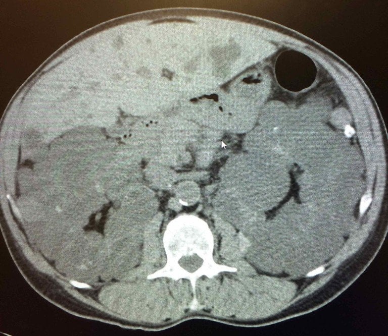
A 37-year-old female presents to her family physician with recurring abdominal and flank pain. She mentions her mother suffered from kidney problems but doesn’t know many details. Examination reveals a blood pressure of 170/110 mmHg and proteinuria is present on dipstick. Laboratory tests show an elevated hematocrit, microalbuminuria, and microscopic hematuria. A CT scan later reveals the findings seen here.Which of the following conditions is most commonly associated with this patient’s likely diagnosis?
A) Cerebral aneurysm
B) Abdominal aortic aneurysm
C) Renal cell carcinoma
D) Ovarian cyst
Find the answer, discuss this case, and more on Figure 1.
Explore cases, quiz yourself, and solve medical mysteries along with thousands of other medical professionals around the world on Figure 1, the free app where doctors expand their clinical knowledge.

the case is ADPKD
and the A is Answer
Adult variant of renal polycystic disease, with associated liver cysts too. Family history of ‘renal problems’ (mother)